Art and science come together in Illustrating Medicine
If you’ve taken classes in biology or medicine, you have undoubtedly come across hundreds of illustrations showing different parts of the human body. What probably did not cross your mind is the fact that someone drew these illustrations by hand, maybe a half-century ago. One of the illustrators could have been Dorothy Foster Chubb or Nancy Joy, who are having their work celebrated in Illustrating Medicine, an exhibit showcasing original artwork created for Grant’s An Atlas Of Anatomy, published in 1943.
The Atlas is a textbook for medical students and professionals worldwide, known for its detailed anatomy illustrations. Since 1943, it has been republished a dozen times and remains one of the most used textbooks in the field. Unlike other anatomy books including Gray’s Anatomy, which organizes the anatomy by system, Grant’s Atlas was one of the first to show the body by region. Readers looking for information about a particular area of the body, such as the skull, would be able to see all the details of the skeletal, muscular and circulatory systems in one chapter or diagram.
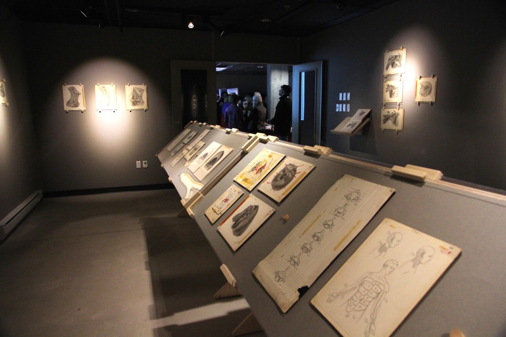
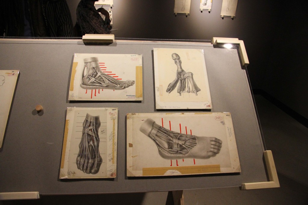
The exhibit is displayed in the same order as the first editions of the atlas, starting with the upper limbs and abdomen, to the pelvis and lower limbs, ending with the vertebrae, head and cranial nerves.
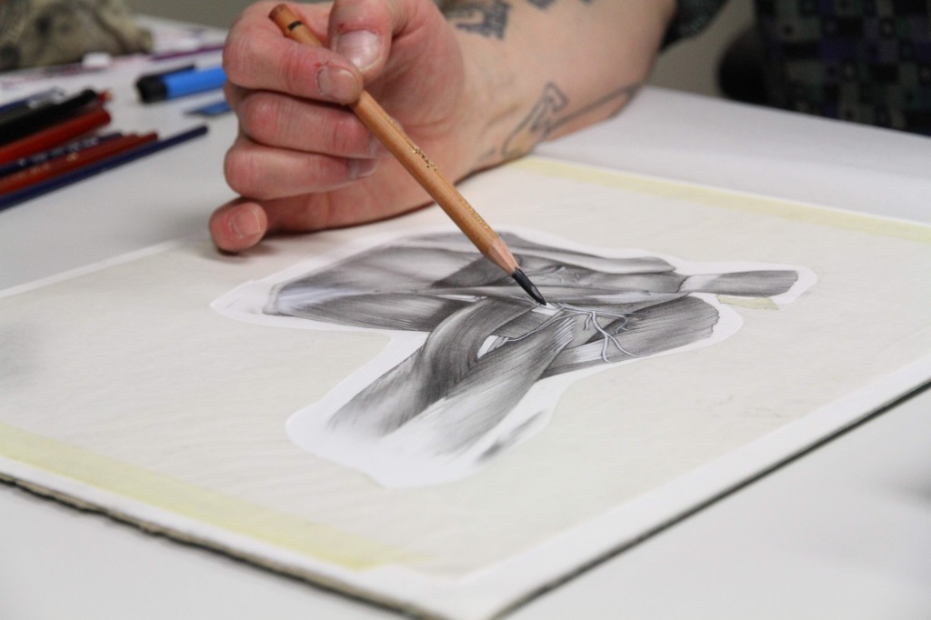
Illustrating Medicine shows a few dozen of Chubb’s and Joy’s original artworks, and a ‘behind-the-scenes’ into their process and collaboration with Dr. Grant.
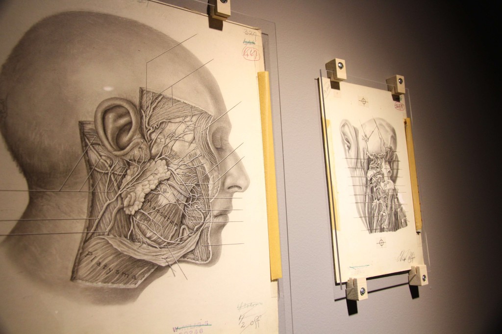
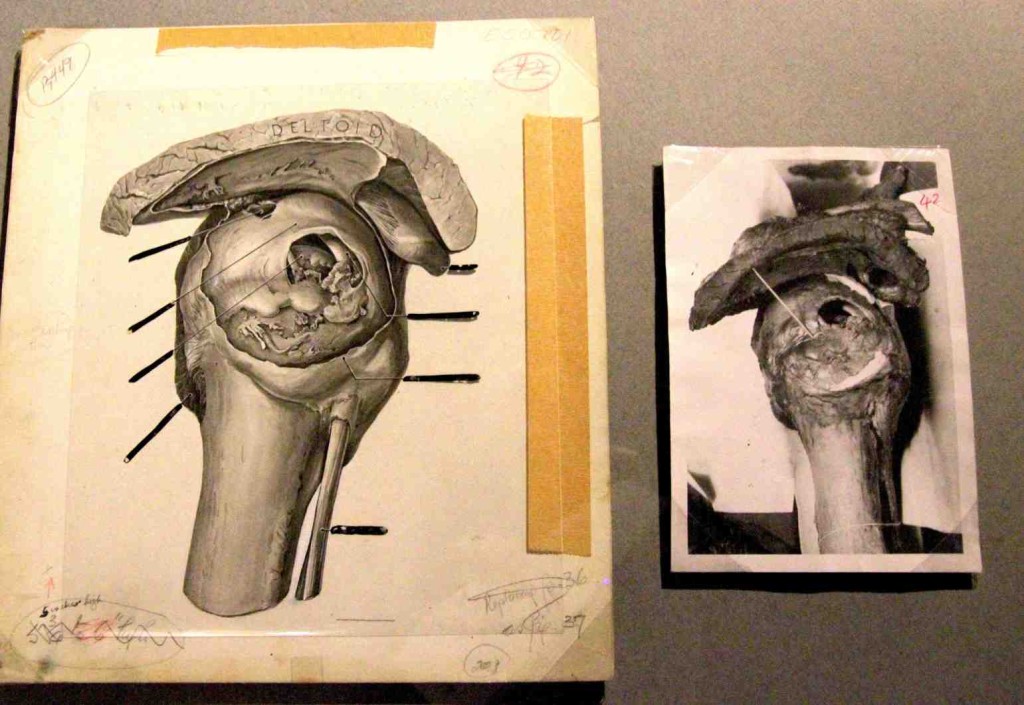
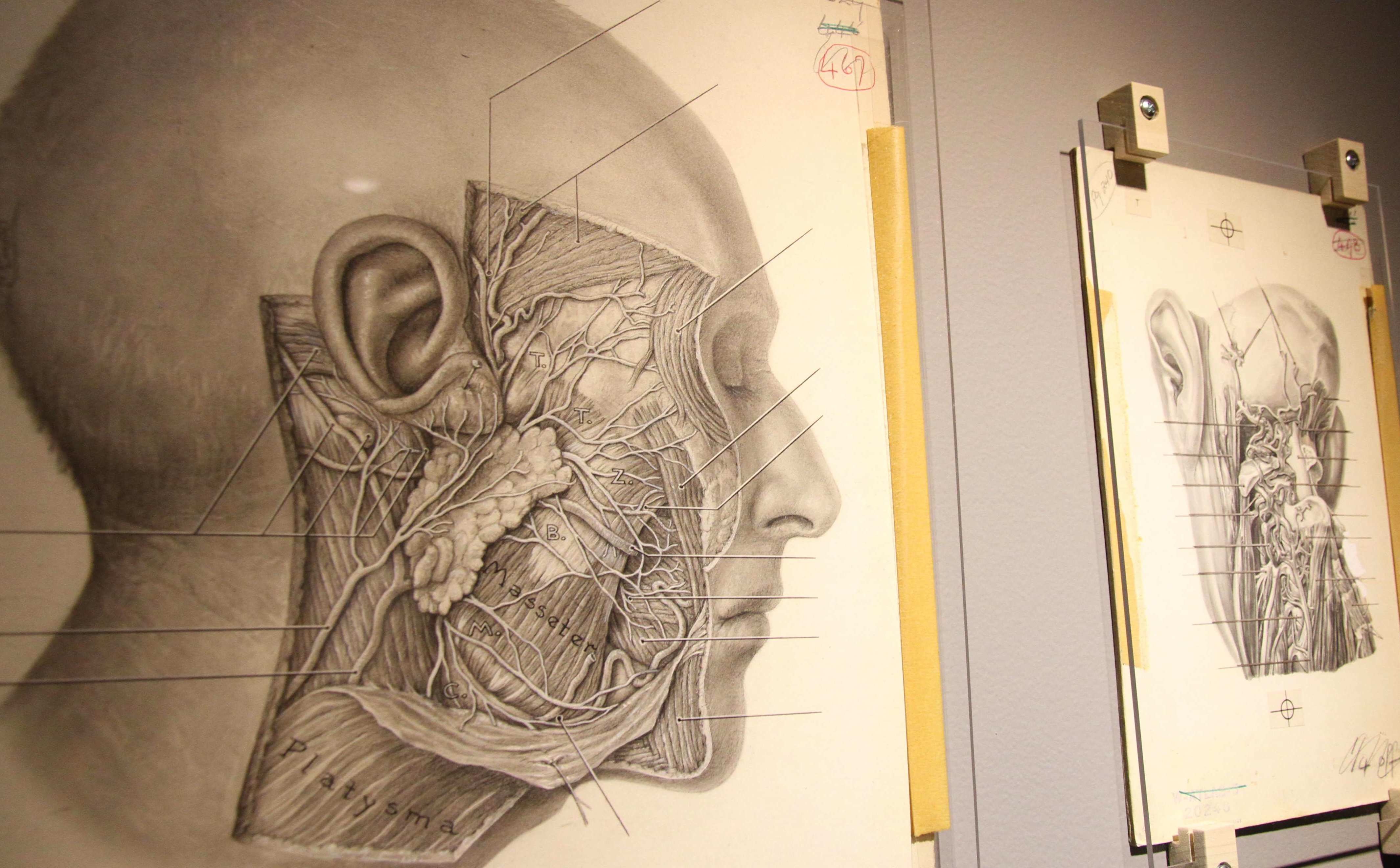
In order to cut costs in production and make the book affordable, Grant’s Atlas did not publish their illustrations with the same amount of detail as the originals. The exhibit is an opportunity to see the works in full detail as they were created, with expert precision and accuracy. Most of the sketches were based on photographs of dissections, which were then traced and consequently made into drawings.
The illustrations were made using different techniques including carbon dust, line drawing and black and white watercolour painting, with each artist sticking to their prefered method. Line drawings often depict bones and venous systems while carbon dust is especially effective for demonstrating muscles and fatty tissues.
Illustrations of anatomy have proved to be more useful than photographs as it allows the reader to see through the different systems. Unlike in a photo, drawings allow you to see the bones, muscles and circulatory system. The artists can play up important elements by using different techniques with highlights and shading. The highlights are key when a specialist is referring to them while performing a dissection.
Looking closely at the drawings, it is difficult to process that someone was able to sketch out intricate details of the human body with such precision. Several illustrations are so realistic that it’s hard not to feel squeamish, but overall they are presented in a way that simply makes you feel in awe of the artists’ talent. Whether or not you are familiar with illustrating, you quickly develop an immense appreciation for the work that went into the drawings and the time it takes to illustrate medicine.
Illustrating Medicine is displayed at the Loyola Campus CJ building Media Gallery until May 1. For more information, visit coms.concordia.ca/illustrating-medicine.html.
Photos by Natasha Taggart